Chapter Twenty-Three: Hypoosmolal States–Hyponatremia
Chapter Twenty-three: Hypoosmolal States–Hyponatremia part 1
Edited by Sophia Ambruso
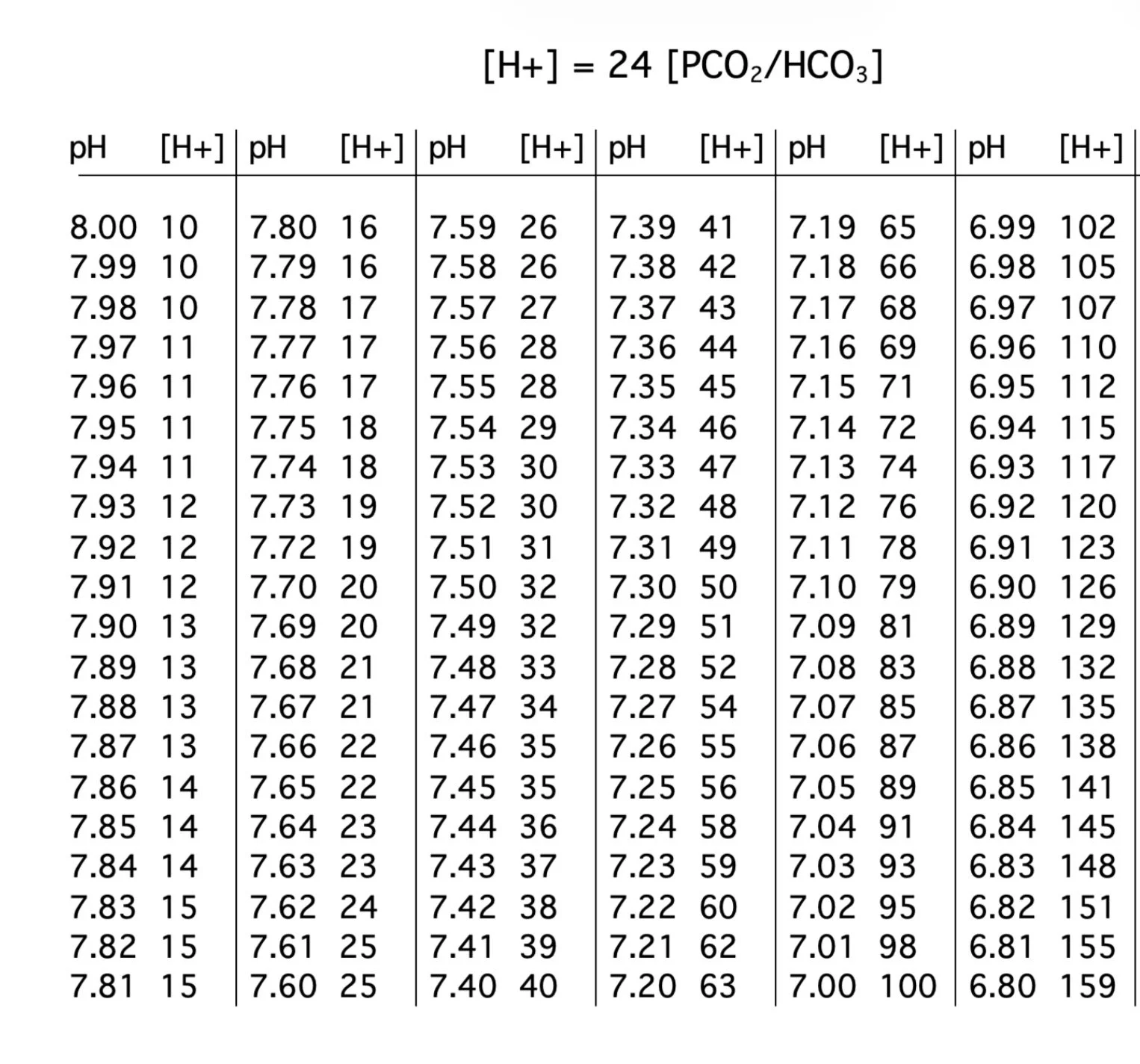
Roger’s Hydrogen to pH table
References
December 19, 2023
Joel had a patient with severe hypothyroidism and hyponatremia- case report
Mel reviewed the endocrine factors that can contribute to hyponatremia in this report (from the 2020 ASN quiz and questionnaire): Pattern Recognition versus Pathogenesis - PMC and amy shared this too: Impact of etiology, age and gender on onset and severity of hyponatremia in patients with hypopituitarism: retrospective analysis in a specialised endocrine unit
JC mentioned reports of sodium loss from biliary sodium as a cause of hyponatremia: Severe Persistent Hyponatremia: A Rare Presentation of Biliary Fluid Loss - PMC , Hyponatremia after Choledochostomy and T Tube Drainage but here’s a reference that did measure biliary sodium concentration in children and found the concentration in bile was similar to plasma (which can lead to significant sodium losses! But not hyponatremia unless replete with dilute fluids ;) 10.1001/archpedi.1986.02140200045024
Here’s a set of articles by Tom Berl on hyponatremia- the first on solute intake and the second in setting of CKD (enjoy! These are really great!): Impact of solute intake on urine flow and water excretion (this figure is brilliant!) and Dysnatremias in Patients With Kidney Disease - PMC Especially enjoy Figure 6! (and check out notes from chapter 9 that review reduced ability to maximally dilute the urine in older individuals- plus here’s a few more great references on the aging kidney: THE AGING KIDNEY: PHYSIOLOGICAL CHANGES - PMC, Urine Concentrating and Diluting Ability During Aging - PMC , Aging and physiological changes of the kidneys including changes in glomerular filtration rate
We quickly referred to the Everest trial of tolvaptan in CHF and Anna shared the reference.
Roger mentioned the weight gain that occurs in hyponatremia and marathon running: this study in the NEJM showed that the risk of hyponatremia was greatest in those with longer race times, more weight gain: https://www.nejm.org/doi/10.1056/NEJMoa043901
Amy’s VOG on Gastric Secretions
Dr. Boyer’s review on his original work on bile salts: https://pmc.ncbi.nlm.nih.gov/articles/PMC4091928/
Gastric secretions
From 1961! https://pubmed.ncbi.nlm.nih.gov/13714620/
From 1959! https://physoc.onlinelibrary.wiley.com/doi/abs/10.1113/expphysiol.1960.sp001428
Secretions (2 book in PDF form)
Basic Concepts of Fluid and Electrolyte Therapy (see page 14) https://www.researchgate.net/publication/369203977_BASIC_CONCEPTS_OF_FLUID_AND_ELECTROLYTE_THERAPY_2nd_Edition
Basic Facts of Body Water and Ions (see page 96) https://link.springer.com/book/10.1007/978-3-662-38375-9
Chapter 23: Hypoosmolar States — Hyponatremia
PATHOPHYSIOLOGY
Plasma Sodium and Plasma Osmolality
Plasma sodium is the main determinant of plasma osmolality.
Hyponatremia = Na <135 mEq/L, which usually reflects hypoosmolality.
Why this matters:
Low plasma osmolality causes water to move into cells.
This cellular overhydration produces the symptoms of hyponatremia, particularly in the brain.
The Basic Mechanism of Hyponatremia
Two questions provide a useful framework:
How do patients develop hyponatremia?
Why do they remain hyponatremic?
Generation of Hyponatremia
Either solute loss (Na or K) or water retention can produce hyponatremia.
However, most fluid losses are approximately isoosmotic to plasma.
Isoosmotic fluid loss by itself is neutral with respect to plasma sodium.
It becomes a problem when the lost fluid is replaced with hypoosmotic fluid.
Thus, hyponatremia is usually fundamentally a disorder of water gain relative to solute.
Hypoosmolality generally cannot develop without water intake.
Perpetuation of Hyponatremia
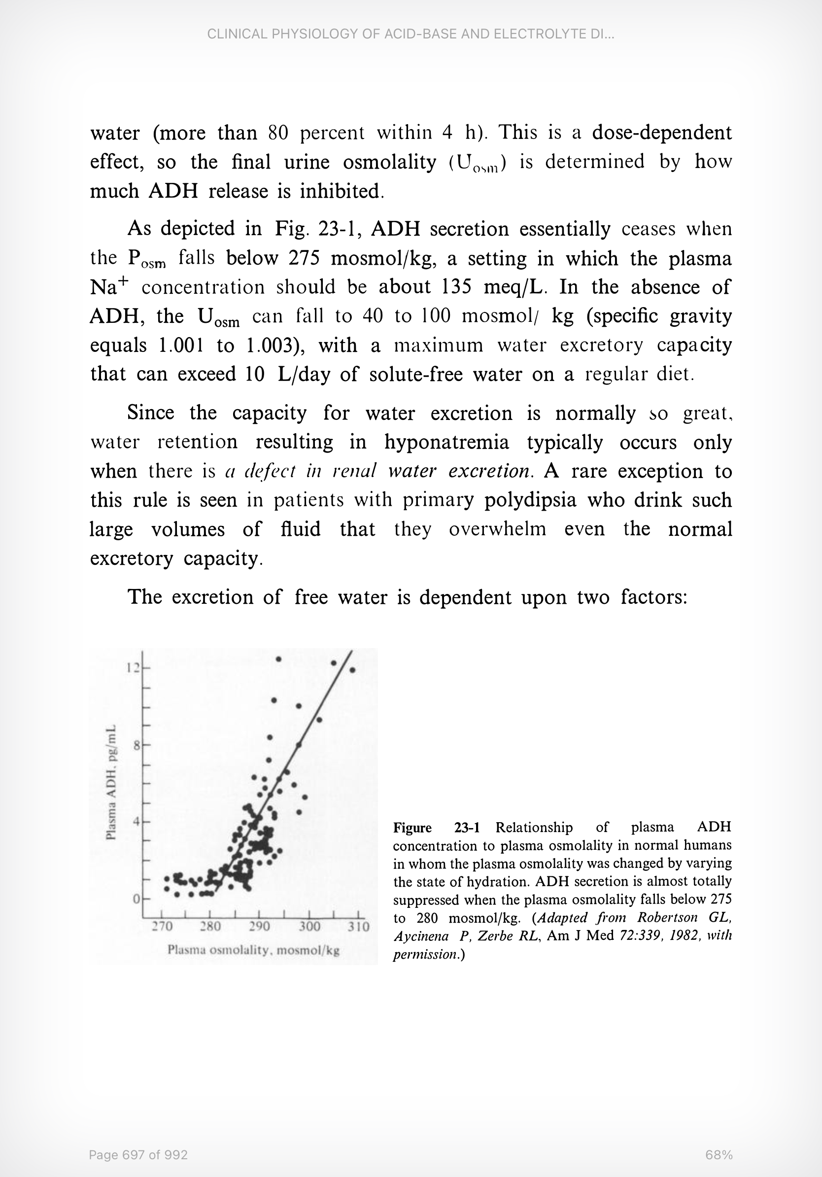
The normal response to falling plasma osmolality is suppression of ADH.
Reduced ADH → dilute urine and increased water excretion.
ADH secretion progressively falls as plasma osmolality falls.
ADH secretion essentially ceases when:
Plasma osmolality falls below approximately 275 mOsm/kg, or
Serum Na approaches 135 mEq/L.
Figure 23-1 appears closer to 280 mOsm/kg.
With maximal ADH suppression:
Urine osmolality falls to approximately 40–100 mOsm/kg H₂O.
Specific gravity falls to approximately 1.001–1.003.
The kidney can potentially excrete >10 L/day of dilute urine.
Because the normal kidney can clear enormous quantities of free water, some defect in renal water excretion is generally required for hyponatremia to persist.
Major exception: primary polydipsia, where water intake overwhelms otherwise intact renal water excretion.
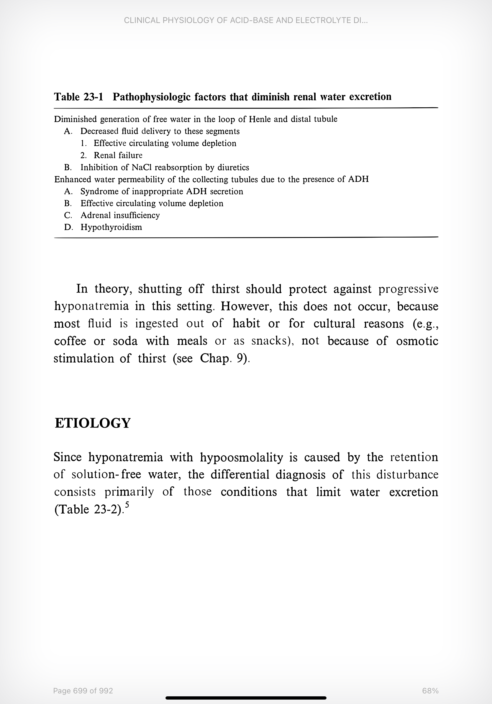
Requirements for Free-Water Excretion
Free-water excretion depends on two processes:
Generation of dilute tubular fluid
NaCl is reabsorbed without water in the diluting segments.
Primarily the thick ascending limb of the loop of Henle.
To a lesser degree, the distal tubule.
See Table 23-1.
Excretion of that dilute fluid
Requires the collecting tubules to remain relatively impermeable to water.
This requires suppression of ADH.
Failure of either process can reduce free-water clearance.
In almost every clinical case, the dominant problem is excess ADH activity, usually from:
SIADH
Decreased effective circulating volume
Important exceptions include:
Oliguric kidney failure
Primary polydipsia
Low-solute intake / “tea and toast” physiology? Not discussed here.
Using Urine Osmolality as an ADH Readout
Urine osmolality provides a functional assessment of ADH activity.
Uosm <100 mOsm/kg: ADH is appropriately suppressed.
Uosm >100 mOsm/kg: ADH effect is present.
In clinically significant ADH-mediated hyponatremia, Uosm is often >300 mOsm/kg.
Thought Experiment: It Doesn’t Take Much
Assume:
Daily solute load = 400 mOsm
Daily water intake = 2 L
If the kidney can dilute urine to 200 mOsm/kg:
400 mOsm ÷ 200 mOsm/kg = 2 L urine
All 2 L of ingested water can be excreted.
But if minimum urine osmolality rises only slightly to 220 mOsm/kg:
400 ÷ 220 = 1.8 L urine
That leaves approximately 200 mL/day of retained water.
Over days or weeks, even this modest impairment can produce progressive hyponatremia.
Why doesn’t suppression of thirst protect the patient?
Because much of human water consumption is habitual, social, or cultural rather than driven strictly by osmotic thirst.
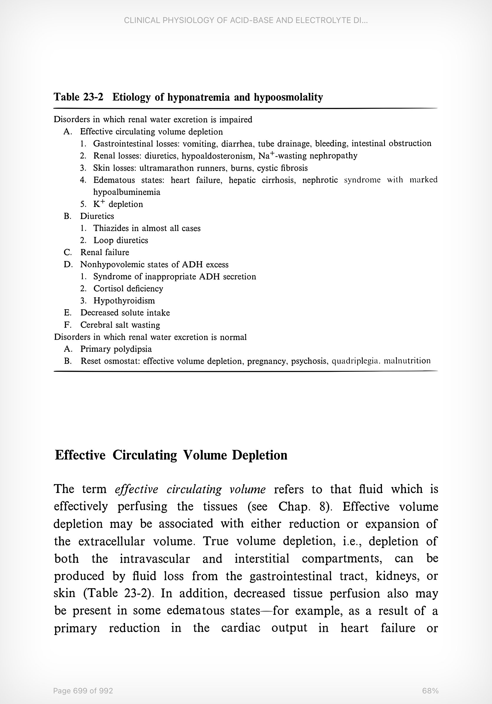
ETIOLOGY
See Table 23-2.
The disorders in which water excretion is impaired are much more important causes of hyponatremia than disorders in which water excretion remains normal.
Effective Circulating Volume Depletion
Effective circulating volume refers to the fluid that is effectively perfusing tissues.
It may be reduced in patients with either decreased or increased total extracellular volume.
True Volume Depletion
Loss of both:
Intravascular fluid
Interstitial fluid
Potential sources:
GI tract
Kidneys
Skin
Other Causes of Reduced Effective Circulating Volume
Decreased vascular resistance
Example: advanced liver disease
Reduced cardiac output
Example: heart failure
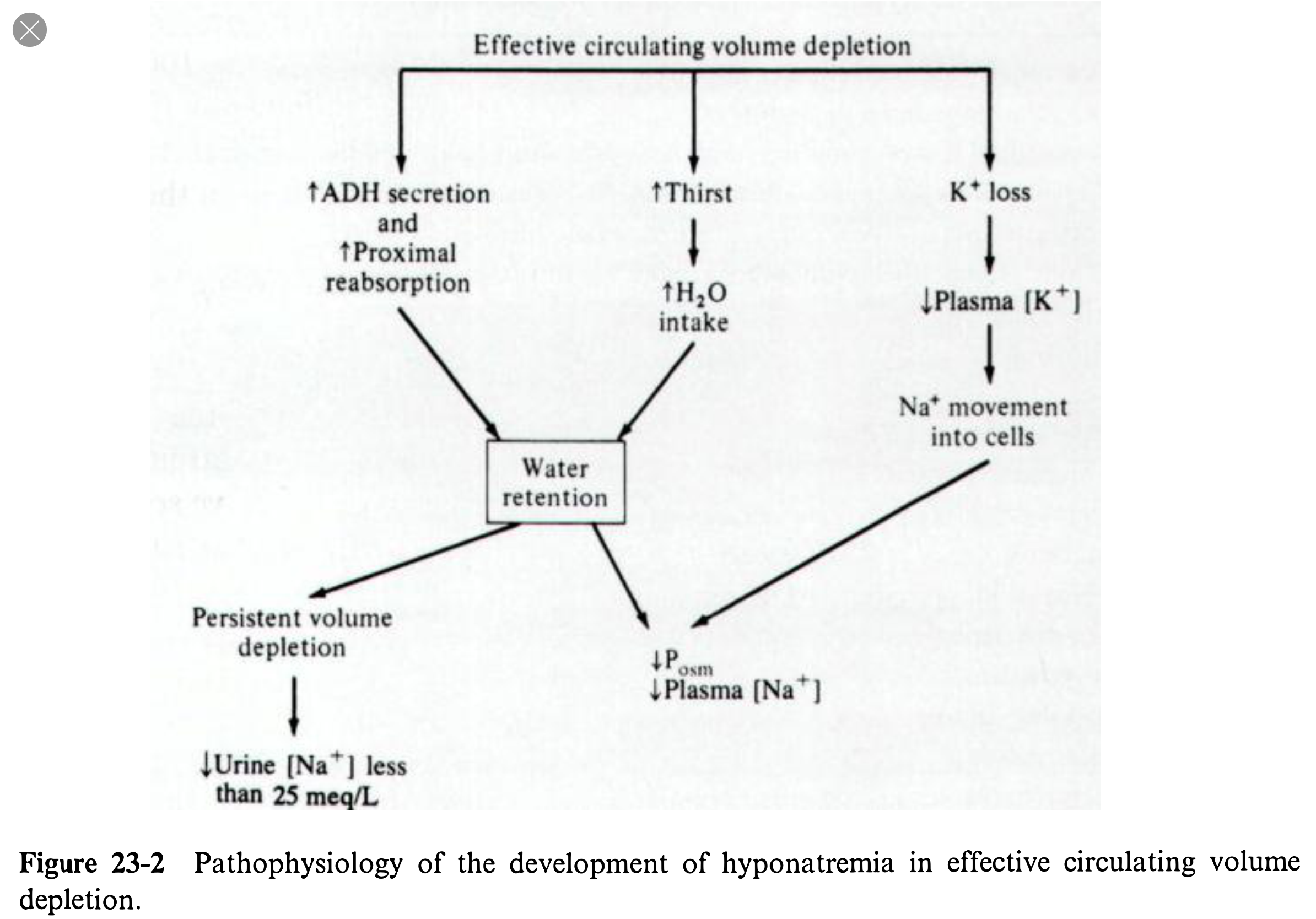
Why Effective Volume Depletion Causes Hyponatremia
Effective volume depletion affects:
Thirst
Potassium balance
Water excretion
ADH
Hypovolemia sensed through arterial baroreceptors is a potent non-osmotic stimulus for ADH secretion.
Reduced Delivery to the Diluting Segments
Volume depletion can also cause:
Reduced GFR
Increased proximal Na reabsorption
Reduced fluid delivery to the diluting segments
This theoretically reduces free-water generation even without ADH.
However, how important is this mechanism?
ADH antagonists can largely reverse impaired water excretion in:
Heart failure
Cirrhosis
Adrenal insufficiency
…without necessarily improving tissue perfusion.
That argues that ADH is doing much of the work.
Severity of Disease Matters
The tendency toward increased ADH and reduced distal delivery increases with the severity of effective volume depletion.
Thus:
Worse heart failure → greater risk of hyponatremia
Worse cirrhosis → greater risk of hyponatremia
Hyponatremia generally does not occur until disease is relatively advanced.
In heart failure, even Na <137 mEq/L is associated with reduced survival.
A seemingly minor reduction in sodium may therefore reflect a major impairment in renal water excretion.
Water Intake Still Matters
The severity of hyponatremia is strongly influenced by how much water the patient consumes.
Ultramarathoners
May lose 10–14 L of sweat.
Sweat contains approximately 20–100 mEq/L of Na + K.
Replacement fluids may contain carbohydrates but relatively little solute.
Symptomatic hyponatremia can develop.
Serum Na may fall below 120 mEq/L.
Cholera
Severe diarrhea from cholera may contain:
Stool Na approximately 120–140 mEq/L.
Low-solute replacement solutions increase the risk of hyponatremia.
Concurrent Potassium Depletion
Potassium depletion can worsen hyponatremia.
K leaves cells to replenish extracellular potassium.
Electroneutrality is partly maintained by Na moving into cells.
This lowers extracellular Na concentration.
Normally, falling plasma osmolality would suppress ADH and permit water excretion.
If ADH remains elevated because of volume depletion, this compensatory mechanism is blocked.
Giving KCl alone can therefore partially correct hyponatremia by reversing the transcellular cation exchange.
Hard to believe Edelman isn’t invoked here.